Low Grade Inflammation in Visceral and Subcutaneous Adipose Tissue Originated from Adipose Derived Stem Cells in Experimental Model of Obesity in Rats under Influence of Pulsed Electromagnetic Field Interaction
Baranowska Agnieszka, Skowron Beata, Ciesielczyk Katarzyna, Guzdek Piotr, Gil Krzysztof, Kaszuba-Zwoińska Jolanta
DOI10.21767/2572-5459.100039
Baranowska Agnieszka1*, Skowron Beata1, Ciesielczyk Katarzyna1, Guzdek Piotr2, Gil Krzysztof 1 and Kaszuba-ZwoiÃÆââ¬Â¦Ãâââ¬Å¾ska Jolanta1
1Department of Pathophysiology, Jagiellonian University Medical College, Cracow, Poland
2Institute of Electron Technology, Cracow, Poland
- *Corresponding Author:
- Baranowska Agnieszka
Department of Pathophysiology
Jagiellonian University Medical College
Cracow, Poland
Tel: +48 12 422 04 11
E-mail: abaranowska@cm-uj.krakow.pl
Received date: January 01, 2018; Accepted date: January 16, 2018; Published date: January 19, 2018
Citation: Agnieszka B (2018) Low Grade Inflammation in Visceral and Subcutaneous Adipose Tissue Originated from Adipose Derived Stem Cells in Experimental Model Of Obesity In Rats Under Influence Of Pulsed Electromagnetic Field Interaction. J Anim Res Nutr Vol No 3: Iss no: 1:1. doi:10.21767/2572-5459.100039
Copyright: © 2018 Agnieszka, et al. This is an open-access article distributed under the terms of the Creative Commons Attribution License, which permits unrestricted use, distribution, and reproduction in any medium, provided the original author and source are credited.
Abstract
Objective: Aim of our study was to examine if pulsed electromagnetic field (PEMF) treatment of the adipose derived stem cells (ADSCs) originating from differently localized adipose tissue, affects the pro-inflammatory cytokines and resistin release dependently on age, gender and used diet.
Methods: Cultures of ADSCs originated from pups and adult animals of both gender grown on low fat ( LF) and high fat (HF) diet for 21 days of obesity inducing time, after which the adipose tissue and blood samples for serum were collected. ADSCs cultures were exposed to PEMF (7 Hz, 30 mT) thrice, 4 h per day. Proinflammatory cytokines and resistin levels were determined by ELISA.
Results: The serum level of TNFα and IL-6 cytokines in HF diet female and male adults was significantly higher as compared to LF diet serum level. HF diet also caused changes in resistin serum level. ADSCs of PEMF exposed LF diet female pups released more TNFα than unexposed ones. IL-6 level in ADSCs cultures of PEMF exposed female pups was higher than in controls. PEMF exposure of ADSCs of both genders of HF diet pups caused increased secretion of TNFα. On the other hand IL-6 release by ADSCs of female pups upon PEMF treatment was diminished. PEMF exposure of ADSCs of both genders of LF diet pups caused increased secretion of resistin. Different results were obtained in case of HF diet pups ADSCs. ADSCs cultures after PEMF exposure originating from male and female adults produced more resistin.
Conclusion: We found that PEMF exposure can modify metabolic activity of ADSCs.
Keywords
Low grade inflammation; Adipose tissue; Pulsed electromagnetic field; Obesity
Introduction
Nowadays low frequency electromagnetic fields are used in the form of magnetotherapy or magnetostimulation as a noninvasive and simple manner to treat a variety of conditions, like Alzheimer's, Parkinson's disease, multiple sclerosis or many diseases with inflammatory patomechanisms, both in medicine and dentistry [1]. The impact of pulsed magnetic field (PMF) is broad and includes anti-inflammatory, antiallodynic character and also and pro-regenerative effect in rodent models [2-6]. The current study revealed that PEMF exposure can modify metabolic activity of ADSCs. Several epidemiologic studies have implicated visceral fat (VAT) as a major risk factor for insulin resistance, type 2 diabetes mellitus, cardiovascular disease, stroke, metabolic syndrome and death [7]. Obesity is an inflammation of the body, showing signs of inflammation at a low level. In addition, it strongly influences the development of insulin resistance and type 2 diabetes [8]. It is well known that high fat containing diet induces obesity and metabolic disorders in rodent experimental studies [9]. Our experiment shows the ontogenic process in animals of both genders including a modified environmental issue - their diet. The research has shown changes in inflammatory cytokines both in serum and in vivo conditions. The consumption of food rich in fat during pregnancy and lactation predisposes to excessive weight gain in later life [10]. Maternal obesity is recognized to trigger stress factors including pro-inflammatory cytokines and oxidative stress molecules [11]. The accumulated knowledge allows for a statement that obesity plays an important role in the pathogenesis of low-grade inflammation related to metabolic disorders [12]. The adipose tissue (AT) is an endocrine organ which affects, among others, the vascular and immune systems and plays an important function in metabolism [13]. One of the AT roles is secretion of adipokines and cytokines which have a signaling character. The above factors play a significant role in maintaining health but from the other side they also participate in pathophysiology of obesity [14]. AT consists of a fraction of white adipose tissue (WAT) and brown adipose tissue (BAT).
Basic role of WAT is collection of triacylglycerols while BAT takes part in heat production [15]. WAT is responsible for production of bioactive molecules – adipokines, which include leptin, resistin, adiponectin, that affect the body homeostasis [16]. The expression of protein substances is related to the localization of two main fat depots: visceral (VAT) and subcutaneous (SAT) adipose tissues. Results related to the level of proinflammatory molecules showed higher levels in VAT, as compared to SAT, in diabetes mellitus type 2 [17]. Most of these adipokines have a proinflammatory character. Scientific reports showed that in fatty tissue: tumor necrosis factor-α (TNFα), interleukin-6 (IL-6), monocyte chemoattractant protein-1 (MCP-1) are elevated [18,19]. Another pro-inflammatory adipokinin is resistin, produced by adipocytes and macrophages [20]. Resistin is also known as adipose tissue-specific secretory factor (ADSF) and was found to be released from adipose tissue and involved in other physiological systems, such as inflammation and energy homeostasis [21-23]. Resitin expression can be upregulated by some interleukins such as IL-6, IL-1 and TNFα as well as by some inflammatory stimuli like lipopolysaccharide (LPS) from Gram negative bacteria. Resistin is primarily related to insulin sensitivity and adipocyte differentiation [24]. In obesity and metabolic syndrome, higher levels of adipokines were observed in blood, like resistin, leptin, adiponectin and also C reactive protein (CRP) [25]. Research has shown that overweight reduces inflammation by decreasing the serum CRP, IL-6 leptin and resistin [26,27]. Shang et al. used ADSCs to supervise disease with inflammatory or autoimmune character, but the correlation between obesity and inflammation remains still unexplained [28]. Thus resistin is reputed to contribute to insulin resistance from one side, and from the other side some research results suggest that resistin may be a link in the well-known association between inflammation and insulin resistance [29]. WNT proteins are family of autocrine and paracrine growth factors that affect biological and developmental processes. They also mediate signal transduction pathways in fat tissue. WNT5A, a noncanonical WNT ligand, has been shown to promote AT inflammation and insulin resistance in animal studies. Elevated non-canonical WNT5A signaling in VAT contributes to the exacerbated IL-6 production in this depot and the low-grade systemic inflammation typically associated with visceral adiposity [30].
Materials and Methods
Animals and ethics statement
Animals were purchased from the central animal house of the Pharmacy Faculty at the Jagiellonian University Medical College (JUMC) in Cracow. All procedures related to laboratory Wistar rats were approved by the Animal Bioethical Committee of Jagiellonian University, Cracow, Poland (resolution no. 84/2014). Dams and adult rats were housed in air conditioned room with constant temperature of 20°C +/- 4 ° C with a 12 hour day/night cycle with humidity level of 55% ±-10%. To ensure that the welfare of the Wistar albino rats was at the highest level possible, we provided lux in an expedition room at 25-50 lux. The air exchange was at the recommended level of 15-20 exchanges per hour. The animals were randomized to an experimental group and received chow and water ad libitum. Female rats, during mating time, pregnancy and lactation (21 days) received the LF or HF diets, respectively. At birth, dams were sexed and randomized in groups with 8 litters. To implement one of the Principles of Humane Experimental Technique, the remaining pups were left to adulthood.
Diet
In our experimental model of obesity, we used the following dietary groups: (1,3) group on low fat (LF) diet, with female or male pups with mother; (2,4) group on high fat (HF) diet, with female or male pups with mother; (5,7) group on LF diet with adult male or female; (6,8) group on HF diet with adult male or female. The LF diet (Labofeed B, Pasze Kcynia, Poland) supplement contained 25% of protein, 8% of fat and 67% of carbohydrates. The obesity-inducing diet, DIO (VERSELE-LAGA Opti Life Adult Active, Belgium) contained: 32% of protein, 22% of fat and 40% of carbohydrates was shown in Table 1.
| Diet component | LF diet | HF diet |
|---|---|---|
| Carbohydrates, ashes and minerals [%] | 67 | 46 |
| Protein [%] | 25 | 32 |
| Fats [%] | 8 | 22 |
| Energy density [kcal/g] | 2.75 | 4.7 |
Table 1: Compositions of 1 gram of low fat diet (LF) versus high fat diet (HF).
Blood collection
After a 21-days period of feeding animals with different diets, all members of the experimental groups were terminally anaesthetized with pentobarbital (Morbital, PuÃÆââ¬Â¦Ãâââ¬Å¡awy, 200 mg per dose). We then collected blood samples from the cardiac vein using standard surgical techniques and centrifugation to obtain serum samples for further analytic procedures.
Isolation and cell culture of ADSCs
ADSCs in female rats were obtained from white adipose tissue from the subcutaneous area. From male rats, we isolated ADSCs from the visceral area according to a published method based on adult BalbC mice [31]. Isolated fat tissues were washed with phosphate-buffered saline (PBS, Sigma-Aldrich, Germany) containing 1% penicillin/streptomycin solution (Sigma-Aldrich, Germany), minced and digested with collagenase type (1 mg/mL; Gibco by Life Technologies, USA) at 37°C for 1 h. Enzyme activity was neutralized with a Dulbecco’s modified eagle’s medium (DMEM, Sigma-Aldrich, Germany) containing 10% FBS (Gibco by Life Technologies, USA) and 1% penicillin/streptomycin solution. Afterwards, the reaction cocktail obtained from disintegrated tissues was filtered (100 μm pore diameter filters, Fisher Scientific, USA), centrifuged at 300 g for 10 min. to obtain a high-density cell pellet. The cell pellet was suspended in DMEM supplemented with 10% fetal bovine serum (FBS; Gibco by Life Technologies, USA) and 1% penicillin/streptomycin solution (Sigma-Aldrich, Germany) and placed in T75 flasks (Corning, Sigma-Aldrich, Germany), incubated overnight at 37°C in a 5% CO2 incubator at 90% humidity. Unattached cells and non-adherent red blood cells were removed after 24 h by washing with antibiotic supplemented PBS. Adherent cells (ADCSs) were suspended in DMEM supplemented with 10% FBS and 1% penicillin/streptomycin solution. The medium was changed at 72 h intervals until the cells became confluent. When the cells reached 80-90% confluence, they were placed in trypsin - EDTA solution (0.25% wt/vol; Sigma-Aldrich, Germany) for 10-15 min. at 37°C, and dissociated by trituration. To arrest the trypsin reaction, DMEM containing 10% FBS and 1% penicillin/streptomycin solution was added the cell suspension and the cells were centrifuged at 416 g for 10 min. ADSCs were suspended in DMEM medium supplemented with 10% FBS and 1% penicillin/streptomycin solution, counted with a hemocytometer and seeded on 96-well plate (Corning, Sigma- Aldrich, Germany) in triplicate at a density of 0.25 × 10⁶ cells/ml per each sample and cultured at 37°C with 5% CO2 and humidified atmosphere.
Magnetic stimulation of ADSCs cultures
PEMF stimulation was started after 24 h of ADSCs culture. A generator produced and kindly provided by the Institute of Electron Technology (Cracow, Poland) generated pulsed electromagnetic field with a frequency 7 Hz at a flux density of 30 mT inside the cell culture incubator. A 96-well plate with ADSC cultures was placed in the generator’s pocket and exposed to EMF for 4 h daily at 24 h intervals, during three consecutive days; this corresponded to a total of three PEMF exposures over the 3-day period. Control samples were placed in the same incubator but at a 35 cm distance from the generator.
Cytokines production
Inflammatory cytokines and resistin levels were measured in blood serum samples obtained from animals of all experimental groups as well as in supernatants originated from ADSCs cultures 24 h after the last PEMF exposure and from unexposed control cultures, respectively. The cytokine concentrations TNFα, IL-6 (Diaclone SAS, France) and resistin (SunredBio, China) were estimated by enzyme-linked immunosorbent assay (ELISA) strictly according to the manufacturer’s procedure.
Statistical analysis
The data were expressed as mean (+) standard deviation (S.D.) and compared using the Student t-test considering P<0.05 defined as significantly different.
Results
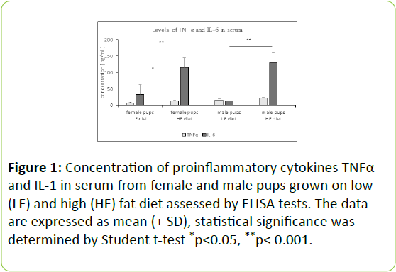
Serum level of proinflammatory cytokines and resistin in rat pups of both genders grown on LF and HF diet: In serum of female and male pups grown on HF diet the level of both proinflammatory cytokines TNFα and IL-6 was increased, but the really big difference was observed in IL-6 level in serum (p<0.02) comparing female and male pups fed with LF diet (Figure 1).
Resistin level in serum of female and male pups grown on HF diet was elevated in comparison to serum resistin level of rats of both genders fed the LF diet (Figure 2).
Serum level of proinflammatory cytokines and resistin in rat adults of both genders grown on LF and HF diet: The serum level of TNFα and IL-6 cytokines in the group of female and male adults grown on HF diet were significantly higher as compared to serum level of these cytokines in rats grown on LF diet (Figure 3).
HF diet caused changes in the serum level of resistin in adult rats of both genders when compared to LF diet; higher content of resistin was measured in serum of adult animals induced by HF diet (Figure 4).
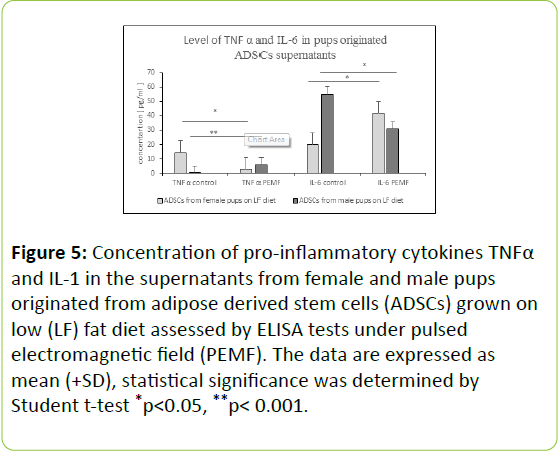
PEMF exposure induced changes in proinflammatory cytokines level in ADSCs cell cultures originated from female and male rat pups grown on LF diet: In in vitro cell culture conditions, PEMF exposure of ADSCs isolated from female and male rat pups grown on LF diet caused changes in proinflammatory cytokines levels in ADSCs cell culture supernatants. PEMF exposed ADSCs cell cultures, originating from female pups, released more TNFα than unexposed ADSCs cell cultures, the opposite effect was obtained in case of male originated, PEMF exposed ADSCs cell cultures (Figure 5).
Figure 5: Concentration of pro-inflammatory cytokines TNFα and IL-1 in the supernatants from female and male pups originated from adipose derived stem cells (ADSCs) grown on low (LF) fat diet assessed by ELISA tests under pulsed electromagnetic field (PEMF). The data are expressed as mean (+SD), statistical significance was determined by Student t-test *p< 0.05, **p< 0.001.
IL-6 level in PEMF exposed ADSCs cell cultures from female pups was higher than in controls not exposed to PEMF, the opposite effect was induced in ADSCs cell cultures from male pups. Upon PEMF treatment there was a significant increase of IL-6 cytokine produced to the milieu (Figure 5).
PEMF exposure induced changes in proinflammatory cytokines level in ADSCs cell cultures originated from female and male rat pups grown on HF diet: PEMF exposure of ADSCs cell cultures originating from pups of both genders grown on HF diet caused increased secretion of TNFα to cell culture medium by ADSCs, as opposed to ADSCs which were not exposed to PEMF. While the release of IL-6 by ADSCs originated from female pups upon PEMF treatment was diminished, ADSCs from male pups exposed to PEMF produced more IL-6 than ADSCs in controls (Figure 6).
Figure 6: Concentration of pro-inflammatory cytokines TNFα and IL-1 in supernatants from female and male pups originated from adipose derived stem cells (ADSCs) grown on HF diet assessed by ELISA tests under pulsed electromagnetic field (PEMF). The data are expressed as mean (+SD), statistical significance was determined by Student t-test *p< 0.05.
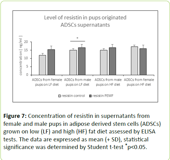
Resistin level changes induced by PEMF exposure in ADSCs cell cultures originated from rat pups and adults grown on HF or LF diet: PEMF treatment of ADSCs cell cultures from pups of both genders fed with LF diet caused increased secretion of resistin cell culture medium by ADSCs as compared to ADSCs cultures which were not treated with PEMF. Different results were obtained in case of ADSCs originated from HF diet pups. ADSCs cell cultures isolated from female pups grown on HF diet have shown slightly diminished resistin level upon PEMF influence (Figure 7).
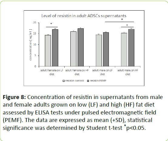
The ADSCs cell cultures from adults of both genders fed with LF and HF diet when treated with PEMF, a slight elevation of the measured resistin content in cell culture supernatants was observed in Figure 8.
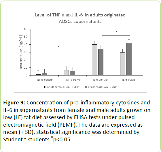
PEMF exposure induced changes in proinflammatory cytokines level in ADSCs cell cultures originated from adult female and male rats grown on LF and HF diet: The level of IL-6 evaluated in ADSCs cell culture supernatants originating from adult females grown on LF diet was lowered under PEMF influence, an opposing effect was observed in ADSCs cell culture supernatants obtained from LF diet raised males, Figure 8.The TNFα level was increased in ADSCs cell cultures supernatants originating from both genders upon PEMF treatment (Figure 8). The ADSCs from male adult on HF diet under PEMF showed in inflammatory profile a significantly higher level of TNFα and IL-6 (Figure 9).
Figure 9: Concentration of pro-inflammatory cytokines and IL-6 in supernatants from female and male adults grown on low (LF) fat diet assessed by ELISA tests under pulsed electromagnetic field (PEMF). The data are expressed as mean (+ SD), statistical significance was determined by Student t-students *p< 0.05.
Spectacular increase of the TNFα amount released to cell culture milieu occurred in ADSCs cell cultures isolated from both genders grown on HF diet, on the other hand when it comes to Il-6 measurements, there was nearly no effect in IL-6 level in ADSCs originated from HF grown females, and a slight increase in ADSCs supernatants obtained from HF diet males (Figure 10).
Figure 10: Concentration of pro-inflammatory cytokines TNFα and IL-6 in supernatants from male and female adults grown on high (HF) fat diet assessed by ELISA tests under pulsed electromagnetic field (PEMF). The data are expressed as mean (+ SD), statistical significance was determined by Student t-students *p< 0.05, **p< 0.001.
Discussion
Maternal obesity caused by high fat diet is responsible for chronic low grade inflammation in progeny placenta, adipose tissue, liver, vascular system and brain [32]. During in utero time period, when the mother received obesity-inducing diet (Neeraj Desai et al.) a higher level of IL-6, TNFα was revealed in fetal plasma [33]. Also TNFα in maternal plasma of animals on HF diet was higher than in controls [34]. The obvious consequence of mother’s diet with high fat levels is systematic inflammation in the offspring. Other studies also support our reports, that in offspring from mothers on HF diet from mating, gestation and lactation, TNFα may be evaluated in 3-weeks-old pups of both genders [35]. In contrast to the above statement, in pregnant rats on HF diet, the maternal plasma IL-6 and TNFα was reduced. These incompatibilities in inflammatory profiles are the results of short exposure to diet induced obesity (DIO) chow compositions and also timing of the harvested tissue [36]. Our findings support the resistin levels evaluated in male offspring from overweight mothers on HF diet in postnatal day (PND) [22,37].
Overweight contributes to collection fat in AT, liver and muscle to cause activation of macrophages by secretion a proinflammatory proteins like TNFα, CRP, MCP-1, leptin, IL-6, resistin [38]. Results of our study confirm earlier finds that exposure to HF diet is connected to higher level of TNFα and IL-6 in adult male rats [39]. Gil et al. state that long term HF diet in adult mice on the degree of pro-inflammatory cytokines (TNF-α, IL-6, and MCP-1) was higher than in control-animals on LF diet [40]. Introduction of HF diet to adult rodent male species resulted in higher concentration of TNFα, just like in our experiment, but also in growth of other pro-inflammatory biomarkers – CRP, IL-17 [41,42]. In obesity models, WAT accumulated excess fatty tissue and released resistin, with levels correlating to DIO and possibly gene–related issues [43,44].
Scores from Muthulakshmi et al. are consistent with our experiment pro-inflammatory markers panel (TNFα, IL-6, resistin) in male mice and rats on HF diet [45]. Our results are also compatible, with the statement that in rodent male model on HF diet, the level of resistin is higher [46,47]. Other studies which also support our work show that in both genders of adult rats, the level of resistin increased on HF diet [48]. Our data are in agreement with Christine et al. research who found that in adult female mice on high fat diet, there were higher levels of inflammatory molecules, including IL-6 [49]. Acute model of high fat diet was in many reports dedicated to males. Milles et al. proved that in the females on HF diet, the level of TNFα was lower as compared to animals on LF diet. The differences may have resulted from the short duration of the diet [50].
Conclusion
Our previous experiments showed PEMF impact on ADSCs obtained from rat pups of both genders on LF diet having protective effects on viability. Contrary, PEMF exerted influence on ADSCs cell culture originating from adult female rats grown on HF diet by inducing high percentage of early apoptotic cells [51]. Unfortunately, there are little data available in the literature concerning the influence of PEMF on proinflammatory markers in ADSCs from adult rats of both genders on LF and HF diets. However, the available publications provide information that PEMF impact on bone marrow cells from adult female rats on standard diet causes a decrease in TNFα and IL-6 [52]. In our study, PEMF treatment of ADSCs from adult female rats on LF diet presented low IL-6 level, however TNFα concentration increased. Incompatibilities in PEMF influence may have resulted from the time exposure of the cells. Other reports showed osteoclastogenesis in adult females under low frequency pulsed electromagnetic field (ELF-PEMF) exposure. In 6 and 8 day of ELF-PEMF stimulation (intensity of 7.5 Hz, tension of 12.2 mV/cm and time 2 h/ day) TNFα concentration grew as compared to controls [53]. Electromagnetic fields used in obese persons, both men and women, lead to high abdominal fat reduction [54]. In obese mice weight and fat loss was reported after activity of 0.5 T electromagnetic fields [55]. In addition, the use of extremely low frequency field (ELF-MF 7.5 Hz, 0.4 T) on human tissue induced an inhibitory effect on obesity. Research studies carried out on mesenchymal stem cells (MSCs) showed a restrictive influence of ELF–MF on adipogenic differentiation [56]. Mengyao et al. proved that PEMF influence promotes myoblast differentiation, obtained from mice in inflammatory and in vitro conditions [57]. Also in human fibroblast like-cells, the level of inflammatory cytokines (IL-1β, TNFα) was lower with PEMF treatment of 2.25 mT with a frequency of 50 Hz [58]. Our earlier experimental studies analyzing the lipid profile in offspring on DIO feed showed high levels of triglycerides (TG), total cholesterol (TC) and low density lipoproteins (LDL) [59]. ADSCs isolated from young female animals grown on HF diet were susceptible for PEMF treatment what decreased release proinflammatory cytokine like IL-6 and metabolic agent – resistin. Adipose derived mesenchymal stem cells isolated and characterized and then osteogenic differentiation of them was investigated after culturing on the surface of Poly (caprolactone) (PCL) scaffold under treatments of pulsed electromagnetic fields (PEMF) [3]. ADSCs-seeded PCL nanofibrous scaffold in combination with PEMF could be a great option for use in bone tissue engineering applications [60].
References
- SieroÃÆââ¬Â¦Ãâââ¬Å¾ A, CieÃÆââ¬Â¦Ãâââ¬Âºlar G, Kawczyk-Kupka A, Biniszkiewicz T, Bilska Urban, et al. (2002) Application of magnetic fields in medicine. Publishing center Augustana.
- Markov MS (2007) Expanding use of pulsed electromagnetic field therapies. Electromagn Biol Med 26: 257-274.
- Kraszewski W, Syrek P, (2010) Magnetotherapy - The use of a magnetic field in treatment and the risks associated with it. 248: 213-228.
- Gherardini L, Ciuti G, Tognarelli S, Cinti C (2014) Searching for the perfect wave: The effect of radiofrequency electromagnetic fields on cells. Int J Mol Sci 15: 5366-5387.
- Pena-Philippides JC, Yang Y, Bragina O, Hagberg S, Nemoto E (2014) Effect of pulsed electromagnetic field (PEMF) on infarct size and inflammation after cerebral ischemia in mice. Transl Stroke Res 5: 491-500.
- Fernandez MI, Watson PJ, Rowbotham DJ (2007) Effect of pulsed magnetic field therapy on pain reported by human volunteers in a laboratory model of acute pain. J Anaesth 99: 266-269.
- 7. Finelli C, Sommella L, Gioia S, La Sala N, Tarantino G ( 2013) Should visceral fat be reduced to increase longevity? Ageing Res Rev 12: 996-1004.
- Tan CP, Hou YH (2014) First evidence for the anti-inflammatory activity of fucoxanthin in high-fat-diet-induced obesity in mice and the antioxidant functions in PC12 cells. Inflammation 37: 443-450.
- Oliveira Junior SA, Dal Pai-Silva M, Martinez PF, Lima-Leopoldo AP, Campos DH (2010) Diet-induced obesity causes metabolic, endocrine and cardiac alterations in spontaneously hypertensive rats. Med Sci Monit 16: 367-373.
- Jessica L, Bolton BS (2014) Developmental programming of brain and behavior by perinatal diet: Focus on inflammatory mechanisms. Clin Neurosciv 16: 307-320.
- Dong M, Zheng Q, Ford SP, Nathanielsz PW, Ren J (2013) Maternal obesity, lipotoxicity and cardiovascular diseases in offspring. J Mol Cell Cardiol 55: 111-116.
- Ouchi N, Parker JL, Lugus JJ, Walsh K (2011) Adipokines in inflammation and metabolic disease. Nat Rev Immunol 11: 85-97.
- Poulos SP, Hausman DB, Hausman GJ (2010) The development and endocrine functions of adipose tissue. Mol Cell Endocrinol 323: 20-34.
- Booth A, Magnuson A, Fouts J, Foster MT (2016) Adipose tissue: An endocrine organ playing a role in metabolic regulation. Horm Mol Biol Clin Investig 26: 25-42.
- Kuryszko J, SÃÆââ¬Â¦Ãâââ¬Å¡awuta P, Sapikowski (2016) Secretory function of adipose tissue. G Pol J Vet Sci 19: 441-446.
- Ohashi K, Shibata R, Murohara T, Ouchi N (2014) Role of anti-inflammatory adipokines in obesity-related diseases. Trends Endocrinol Metab 25: 348-355.
- Samaras K, Botelho NK, Chisholm DJ, Lord RV (2010) Subcutaneous and visceral adipose tissue gene expression of serum adipokines that predict type 2 diabetes. Obesity 18: 884-889
- Khosravi R, Ka K, Huang T, Khalili S, Nguyen BH, et al. (2013) Tumor necrosis factor- α and interleukin-6: Potential interorgan inflammatory mediators contributing to destructive periodontal disease in obesity or metabolic syndrome. Mediators Inflamm 2013: 728987
- Yudkin JS (2003) Adipose tissue, insulin action and vascular disease: Inflammatory signals. Int J Obes Relat Metab Disord 27: 325-328.
- Schäffler A, Müller-Ladner U, Schölmerich J, Büchler C (2006) Role of adipose tissue as an inflammatory organ in human diseases. Endocr Rev 27: 449-467.
- Adeghate E (2004) An update on the biology and physiology of resistin. Cell Mol Life Sci 61: 2485-2496.
- Stumvoll M, Häring H (2002) Resistin and adiponectin of mice and men. Obes Res 10: 1197-1199.
- Vendrell J, Broch M, Vilarrasa N, Molina A, Gómez JM, et al. (2004) Resistin, adiponectin, ghrelin, leptin, and proinflammatory cytokines: Relationships in obesity. Obes Res 12: 962-971.
- Roumaud P, Martin LJ, (2015) Another pro-inflammatory adipokinin is the resistin produced by adipocytes and macrophages. Roles of leptin, adiponectin and resistin in the transcriptional regulation of steroidogenic genes contributing to decreased Leydig cells function in obesity. Horm Mol Biol Clin Investig 24: 25-45.
- Khosravi R, Ka K, Huang T, Khalili S, Nguyen BH (2005) Adipose tissue, inflammation, and cardiovascular disease. Circ Res 96: 939-949.
- Esposito K, Pontillo A, Di Palo C, Giugliano G, Masella M (2003) Effect of weight loss and lifestyle changes on vascular inflammatory markers in obese women: A randomized trial. JAMA 289: 1799-1804.
- Lee IS, Shin G, Choue R (2010) Shifts in diet from high fat to high carbohydrate improved levels of adipokines and pro-inflammatory cytokines in mice fed a high-fat diet. Endocr J 57: 39-50.
- Wellen KE, Hotamisligil GS (2005) Inflammation, stress, and diabetes. J Clin Invest 115: 1111-1119.
- Shang Q, Bai Y, Wang G, Song Q, Guo C, et al. (2015) Delivery of adipose-derived stem cells attenuates adipose tissue inflammation and insulin resistance in obese mice through remodeling macrophage phenotypes. Stem Cells Dev 24: 2052-2064.
- Zuriaga MA, Fuster JJ, Farb MG, MacLauchlan S, Bretón-Romero R, et al. (2017) Activation of non-canonical WNT signaling in human visceral adipose tissue contributes to local and systemic inflammation. Sci Rep 7: 17326.
- Safford KM, Hicok KC, Safford SD, Halvorsen YD, Wilkison WO, et al. (2002) Neurogenic differentiation of murine and human adipose-derived stromal Cells. Biochem Biophys Res Commun 294: 371-379.
- Dan Z, Yuan XP (2015) Pathophysiological basis for compromised health beyond generations: role of maternal high-fat diet and low-grade chronic inflammation. J Nutr Biochem 26: 1-8.
- Desai N, Roman A, Rochelson B, Gupta M, Xue X, et al. (2013) Maternal metformin treatment decreases fetal inflammation in a rat model of obesity and metabolic syndrome. Am J Obstet Gynecol 209: 136-139.
- Murabayashi N, Sugiyama T, Zhang L, Kamimoto Y, Umekawa T, et al. (2013) Maternal high-fat diets cause insulin resistance through inflammatory changes in fetal adipose tissue. J Obstet Gynecol Reprod Biol 169: 39-44.
- Segovia SA, Vickers MH, Zhang XD, Gray C, Reynolds CM (2015) Maternal supplementation with conjugated linoleic acid in the setting of diet-induced obesity normalises the inflammatory phenotype in mothers and reverses metabolic dysfunction and impaired insulin sensitivity in offspring. Nutr Biochem 26: 1448-1457.
- Crew RC, Waddell BJ, Mark PJ (2016) Maternal obesity induced by a 'cafeteria' diet in the rat does not increase inflammation in maternal, placental or fetal tissues in late gestation. Placenta 39: 33-40
- Shankar K, Kang P, Harrell A, Zhong Y, Marecki JC, et al. (2010) Maternal overweight programs insulin and adiponectin signaling in the offspring. Endocrinology 151: 2577-2589.
- Weisberg SP, McCann D, Desai M, Rosenbaum M, Leibel RL (2003) Obesity is associated with macrophage accumulation in adipose tissue. Clin Invest 112: 1796-1808.
- Ghiasi R, Ghadiri SF, Mohaddes G, Alihemmati A, Somi MH, et al. (2016) Influance of regular swimming on serum levels of CRP, IL-6, TNF-α in high-fat diet-induced type 2 diabetic rats. Physiol Biophys 35: 469-476.
- Gil HW, Lee EY, Lee JH, Kim YS, Lee BE, et al. (2015) Dioscorea batatas extract attenuates high-fat diet-induced obesity in mice by decreasing expression of inflammatory cytokines. Med Sci Monit 21: 489-495.
- Maithilikarpagaselvi N, Sridhar MG, Swaminathan RP, Sripradha R (2016) Preventive effect of curcumin on inflammation, oxidative stress and insulin resistance in high-fat fed obese rats. Complement Integr Med 13: 137-143.
- DeClercq VC, Goldsby JS, McMurray DN, Chapkin RS (2016) Distinct adipose depots from mice differentially respond to a high-fat, high-salt diet. J Nutr 146: 1189-1196.
- Lu H, Wang H, Wen Y, Zhang M, Lin H (2006) Roles of adipocyte derived hormone adiponectin and resistin in insulin resistance of type 2 diabetes. World J Gastroenterol 12: 1747-1751.
- Bokarewa M, Nagev I, Dahlberg L, Smith U, Tarkowski A (2005) Resistin, an adipokine with potentproinflammatory properties. J Immunol 174: 5789-5795.
- Muthulakshmi (2015) Gene expression profile of high-fat diet-fed C57BL/6J mice: in search of potential role of azelaic acid. J Physiol Biochem 71: 29-42.
- Steppan CM, Bailey ST, Bhat S, Brown EJ, Banerjee RR, et al. (2001) The hormone resistin links obesity to diabetes. Nature 409: 307-312.
- Aborehab NM, Bishbishy MH, Waly NE (2016) Resistin mediates tomato and broccoli extract effects on glucose homeostasis in high fat diet-induced obesity in rats. BMC Complement Altern Med 16: 225.
- Szulinska M, Musialik K, Suliburska J, Lis I, Bogdanski P (2014) The effect of L-arginine supplementation on serum resistin concentration in insulin resistance in animal models. Eur Rev Med Pharmacol Sci 18: 575-580.
- Kim CH, Mitchell JB, Bursill CA, Sowers AL, Thetford A, et al. (2015) The nitroxide radical TEMPOL prevents obesity, hyperlipidaemia, elevation of inflammatory cytokines, and modulates atherosclerotic plaque composition in apoE-/-mice. Atherosclerosis 240: 234-241
- Miller CN, Morton HP, Cooney PT, Winters TG, Ramseur KR, et al. ( 2014) Acute exposure to high-fat diets increases hepatic expression of genes related to cell repair and remodeling in female rats. Nutr Res 34: 9385-9349.
- Baranowska A, Skowron B, Nowak B, Ciesielczyk K, Guzdek P, et al. (2017) Changes in viability of rat adipose-derived stem cells isolated from abdominal/perinuclear adipose tissue stimulated with pulsed electromagnetic field. JPhysiol Pharmacol 68: 253-264.
- Chang K, Hong-Shong Chang W, Yu YH, Shih C (2004) Pulsed electromagnetic field stimulation of bone marrow cells derived from ovariectomized rats affects osteoclast formation and local factor production. Bioelectromagnetics 25: 134-141.
- Chang K, Chang WH, Wu ML, Shih C (2003) Effects of different intensities of extremely low frequency pulsed electromagnetic fields on formation of osteoclast-like cells. Bioelectromagnetics 24: 431-439.
- Beilin G, Benech P, Courie R, Benichoux F (2012) Electromagnetic fields applied to the reduction of abdominal obesity. J Cosmet Laser Ther 14: 24-42.
- Nichols TW (2012) Mitochondria of mice and men: moderate magnetic fields in obesity and fatty liver. Med Hypotheses 79: 287-293.
- Leilei D, Hongye F, Huishuang M, Guangfeng Zhao, Yayi H (2014) Extremely low frequency magnetic fields inhibit adipogenesis of human mesenchymal stem cells. Bioelectromagnetics 35: 519-530.
- Mengyao L, Carlin L, Dominigue L, Nianli Z, Waldorff E, et all. (2017) Role of pulsed electromagnetic fields (PEMF) on tenocytes and myoblasts-potential application for treating rotator cuff tears. J Orthop Res 956-964.
- Gómez-Ochoa I, Gómez-Ochoa P, Gómez-Casal F, Cativiela E, Larrad-Mur L, et al. (2011) Pulsed electromagnetic fields decrease proinflammatory cytokine secretion (IL-1β and TNF-α) on human fibroblast-like cell culture. Pulsed electromagnetic fields decrease proinflammatory cytokine secretion (IL-1β and TNF-α) on human fibroblast-like cell culture. Rheumatol Int 1283-1289.
- Baranowska A, Skowron B, Ciesielczyk K, DomagaÃÆââ¬Â¦Ãâââ¬Å¡a J, Thor PJ (2016) Experimental gender related obesity effect of diet. Folia Med Cracov 56: 49-60.
- Arjmand M, Ardeshirylajimi A, Maghsoudi H, Azadian E (2018) Osteogenic differentiation potential of mesenchymal stem cells cultured on nanofibrous scaffold improved in the presence of pulsed electromagnetic field. J Cell Physiol 233: 1061-1070.

Open Access Journals
- Aquaculture & Veterinary Science
- Chemistry & Chemical Sciences
- Clinical Sciences
- Engineering
- General Science
- Genetics & Molecular Biology
- Health Care & Nursing
- Immunology & Microbiology
- Materials Science
- Mathematics & Physics
- Medical Sciences
- Neurology & Psychiatry
- Oncology & Cancer Science
- Pharmaceutical Sciences
